An intramedullary nail is a metal rod placed inside the central canal of a long bone to stabilize certain fractures. Surgeons commonly use one for fractures of the femur or tibia, and sometimes the humerus. Small locking screws near the bone’s ends help control rotation and maintain length. The nail sits within the bone rather than along its outer surface. That position can help share forces while the fracture heals.
Orthopedic trauma researcher Peter V. Giannoudis has published extensively on fracture fixation. A fair paraphrase of a principle reflected in this work is: “Choose fixation to suit the fracture and the patient.” This is a paraphrase, not a verified verbatim quotation. The distinction matters. An intramedullary nail is not suitable for every fracture, and the decision depends on factors such as fracture location, bone quality, alignment, and overall health. A surgeon may assess X-rays or other imaging, then discuss possible benefits and risks. Picture a broken shin bone held in line by a slim rod, with screws visible near the knee and ankle on an X-ray. The image is simple; the planning is not. Recovery also varies, and a nail does not guarantee a quick return to normal activity. Understanding what the device does—and what it cannot do—helps patients ask clearer questions before treatment.
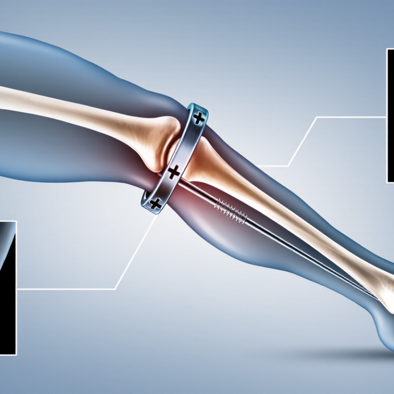
An intramedullary nail is a metal rod placed inside the medullary canal, the hollow center of a long bone. Surgeons commonly use one to stabilize fractures of the femur or tibia, and sometimes the humerus. The rod sits within the bone rather than along its outer surface. That position can help maintain alignment while the fracture heals.
During surgery, the nail is guided into the bone through a small opening near one end. Screws may pass through the bone and nail to limit twisting or shortening. The exact approach depends on the fracture’s location and shape. Imaging helps the surgical team check placement. Materials often include titanium or stainless steel, though the choice varies.
The name can be misleading. It is not a simple nail driven into wood, and it does not make a broken bone heal by itself. It provides internal support while the body repairs the fracture. Some patients may bear weight sooner than others, but that depends on the injury and the surgeon’s instructions. Not every fracture suits this method. Bone quality, nearby joints, and the patient’s overall health all matter. Recovery can be uneven, too; a stable-looking image does not always mean the person feels ready to move normally.

An intramedullary nail is a metal rod placed inside the hollow center of a long bone. It is commonly used to stabilize fractures of the femoral shaft, the tibial shaft, and, in selected cases, the humeral shaft. The nail sits within the bone rather than alongside it, and screws near its ends help control rotation and length. A thigh fracture may need a long nail; a shin fracture often needs a different length and shape.
These nails are especially useful for fractures through the middle portion of a bone, including some displaced or fragmented breaks. Certain fractures near the hip, knee, or ankle can also be treated with specially designed nails, but their location and shape matter. A fracture that extends into a joint may need another approach to align its small joint-surface fragments precisely. Not every long-bone fracture fits a nail.
The decision depends on X-rays, the fracture pattern, bone quality, and the person’s overall condition. Open fractures may sometimes be nailed after careful wound treatment, but infection risk and tissue damage affect the plan. It is easy to hear “long-bone fracture” and assume one device works for all. It doesn’t. Recovery and weight-bearing instructions vary, too.
An intramedullary nail is a metal rod placed inside the hollow center of a long bone, often the femur or tibia. It helps hold broken bone sections aligned while the body repairs them. It is not a clamp. Screws through the bone and nail limit rotation and shortening, while the nail shares some load during movement. This internal support can allow carefully guided weight-bearing, depending on the fracture and the surgeon’s instructions.
The nail sits close to the bone’s central axis, helping resist bending forces. That matters when a person stands or takes a step. A multicentre randomized trial reported in the Journal of Bone and Joint Surgery followed 1,226 patients with tibial shaft fractures. It found no significant difference in its primary outcome between reamed and unreamed nailing approaches. The finding concerns two techniques, not a guarantee of healing. Fracture shape, blood supply, soft-tissue injury, and health all matter. Healing still varies. The best implant position is not a universal recipe, either.
Tips: Follow your weight-bearing instructions, keep wound checks, and report worsening pain, fever, or new numbness promptly. Ask when follow-up imaging is planned. A nail stabilizes the fracture; it does not replace careful recovery.
An intramedullary nail sits inside the hollow center of a long bone, helping hold broken sections in alignment while they heal. It is commonly used for fractures of the thighbone or shinbone. Placement usually happens under general or spinal anesthesia, with X-ray imaging guiding the surgeon. The incision is often near the hip or knee, depending on the bone and fracture. The American Academy of Orthopaedic Surgeons reports that more than 300,000 adults aged 65 and older are hospitalized for hip fractures each year in the United States; that figure shows the scale of fracture care, not the number treated with nails.
A guidewire is passed through the bone’s central canal and across the fracture. The surgeon may widen the canal, then slide the nail over the wire. Small incisions. Steady imaging. The broken ends must be lined up before the nail is secured with screws near both ends. These screws help limit twisting and shortening. The team checks alignment and screw position with live X-rays before closing the wounds. The sequence can look tidy on a diagram; real fractures may be less cooperative, and the exact approach depends on the break’s location and shape. AAOS OrthoInfo and the AO Surgery Reference describe these principles, but only the treating surgical team can explain the plan for an individual fracture.
After intramedullary nailing, recovery depends on the fracture, surrounding tissue, and overall health. Early walking may be encouraged, but weight-bearing limits vary. Follow the surgeon’s plan rather than testing the leg by pain alone. Physical therapy often focuses on safe transfers, knee and ankle motion, and gradually rebuilding strength. Small gains count. Follow-up appointments usually include wound checks and X-rays to assess alignment and healing. A calendar estimate cannot show whether a particular fracture is ready for more activity.
Complications need prompt attention. The SPRINT randomized trial of tibial-shaft fractures reported a reoperation outcome in roughly one in eight patients within a year; this does not mean every patient had a serious complication (SPRINT Investigators, Journal of Bone and Joint Surgery, 2008). Possible problems include infection, delayed healing, nonunion, or irritation from the nail or screws. Contact the care team about increasing pain, spreading redness, drainage, fever, or new difficulty bearing weight. Sudden shortness of breath or chest pain requires emergency care. Even routine recovery can feel uneven, and setbacks deserve reassessment rather than guesswork.
Please visit our Flavor List for inspiration and contact Custom Flavors to discuss your specific flavor needs.
© 2025 Custom Flavors. All Rights Reserved.

"*" indicates required fields
